| Dr. Masahiro Miura's Page Department of Ophthalmology Tokyo Medical University, Ibaraki Medical Center |
|||
| Scanning Laser Ophthalmoscope | |||
| About Miura CV Peer-reviewd paper SLO imaging OCT imaging |
Dr. Miura is collaborating with Ann E Elsner’s labo at the school of Optometry in Indiana University since 2002 for the clinical
application of the polarization-sensitive scanning laser ophhalmoscope.
This collaboration study developed following novel imaging technique. |
||
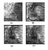 High-contrast retinal vascular images in the eyes with epiretinal membrane. Retinal images of 70 year-old Japanese man with epiretinal membrane. (a) 514 nm scanning laser ophthalmoscope image, (b) depolarized light images, (c) average reflectance image, (d) parallel polarized light image. The epiretinal membrane could be clearly visualized as highly reflected area in the scanning laser ophthalmoscope image at 514 nm. In the depolarized light image, the retinal vessels could be clearly visualized in the area of epiretinal membrane. In the average reflectance image and parallel polarized light image, the retinal vessels were somewhat obscured by epiretinal membrane. (Miura M, Elsner AE, et al (2007) Imaging polarimetry and retinal blood vessel quantification at the epiretinal membrane. J Opt Soc Am A Opt Image Sci Vis 24: 1431-1437.) |
|||
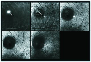 Non-invasive detection of leakage point in central serous chorioretinopathy. Left eye of a 53 year-old-man. Fluorescein angiogram (top left) demonstrating dye leakage near the macula. A depolarized light image (top right) clearly showing the leakage point as a bright dot (white arrow) accompanied with multiple bright dots. In the birefringence image (bottom left), parallel polarized light image (bottom center), or average reflectance image (bottom right), the leakage area could be hardly detected. (Miura M, Elsner, et al (2005) Imaging polarimetry in central serous chorioretinopathy. Am J Ophthalmol 140: 1014-1019.) |
|||
| Depatment of Ophthalmology, Tokyo Medical University, Ibaraki Medical Center (in Japanese) |
|||